
Axiom Bone Level implants (Oct 21)

-
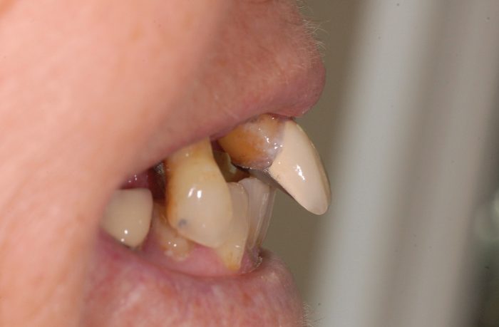 Figure 1: Profile view
 -
 Figure 2: Intra-oral view of remaining maxillary teeth
 -
-
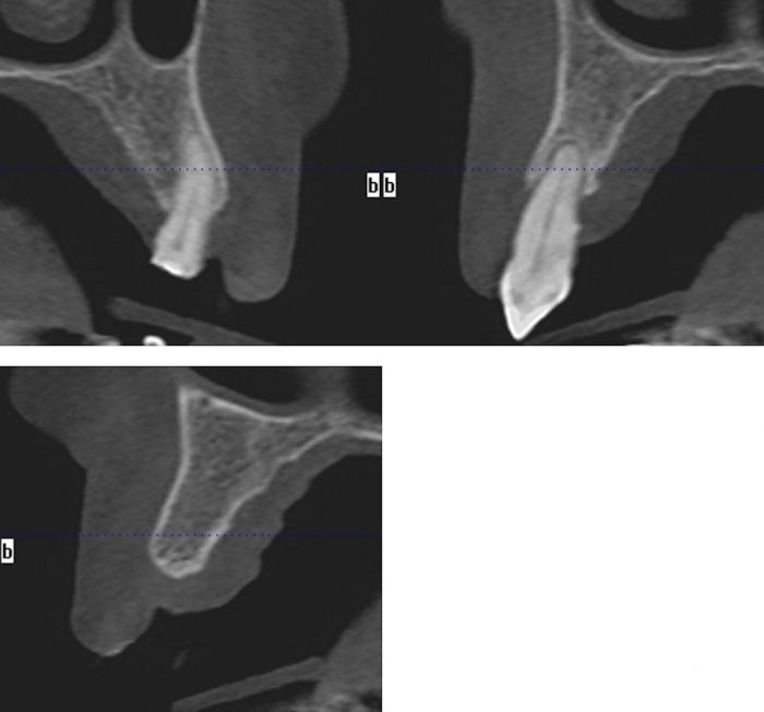 Figure 4: A CBCT scan of the upper jaw was taken to accurately assess the anatomy, bone volume and trajectory of the alveolar ridges
 -
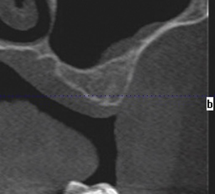 Figure 5: Inadequate bone below the maxillary sinuses
 -
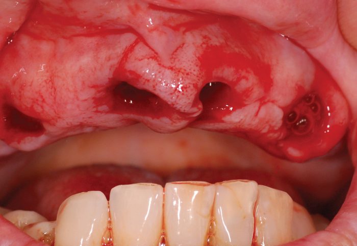 Figure 6: Immediate extraction sockets
 -
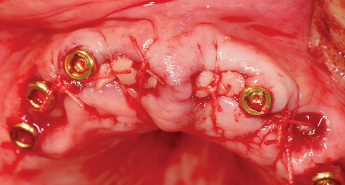 Figure 7: Implants placed, and Locator abutments attached
 -
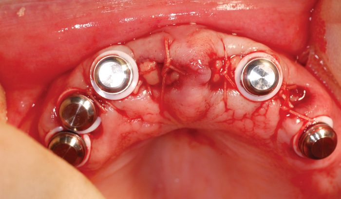 Figure 8: Metal housing on Locator abutments just before pick-up in the denture
 -
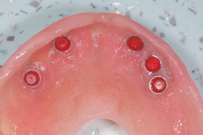 Figure 9: Metal housing precisely secured in the denture
 -
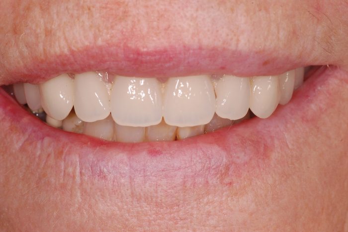 Figure 10: Denture fitted immediately following implant placement
 -
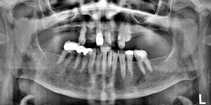
 Figure 3: The OPG radiograph confirmed the absence of any pathology in both jaws and presence of large maxillary sinuses
 -
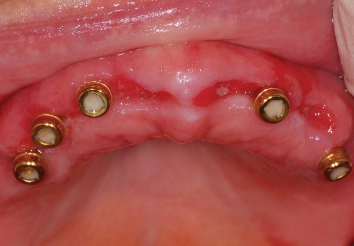 Figure 12: Following three weeks of undisturbed healing
 -
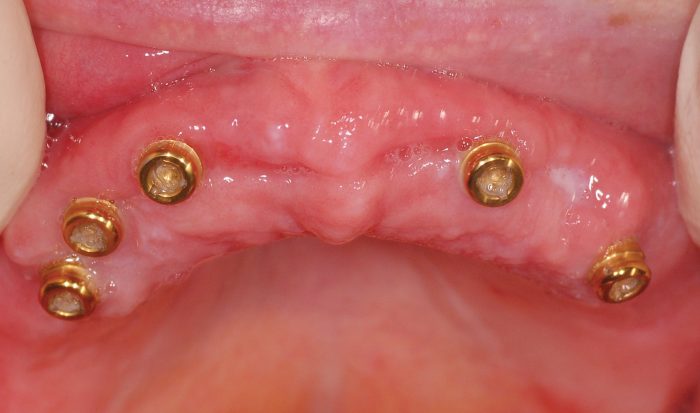 Figure 13: Following 12 weeks of complete healing
 -
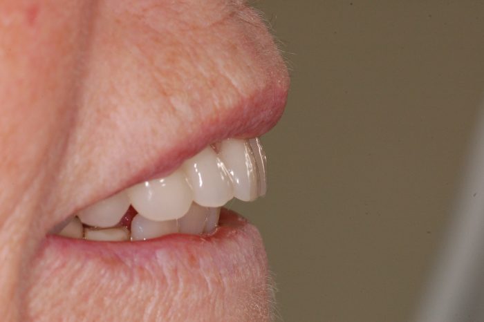 Figure 14: Profile view immediately following implant treatment